Electron Microscopy Uses Which Type Light for Illumination
An online Interactive look at Microscopy The virtual microscope allows people all over the world to study specimens when ever and however they want. Use the same morphological clues for electron microscopy as are used for light microscopy eg.

Transmission Electron Microscope Tem Introduction To Jeol Products Jeol Ltd
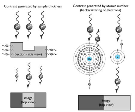
Since the contrast in the electron microscope depends primarily on the differences in the electron density of the organic molecules in the cell the efficiency of a stain is determined by the atomic weight of the stain attached to the biological structures.
. Transmission electron microscope TEM scanning electron microscope SEM etc. These microscopes emit electron beams not light beams toward targets to magnify them. Originally microscopy was based on the use of the light microscope and could provide specimen resolutions on the order of 02 microns.
A small opaque disk about 1 cm in diameter is placed between the illuminator and the condenser lens. Due to the Abbe diffraction limit the spatial resolution of the fluorescence microscope is limited to about half the wavelength of incident light With the development of microscopy super-resolution fluorescence microscopy method provides the possibility for the breakthrough of the diffraction limit such. Slámová M Očenášek V Vander Voort G.
A darkfield microscope is a brightfield microscope that has a small but significant modification to the condenser. To achieve higher resolutions an electron source is required instead of light as the illumination source which allows for resolutions of about 25 Angstroms. Fluorescence microscopy uses intense levels of near-monochromatic illumination and therefore requires one of four main types of lamp.
Examine the sample preferably in the glass vial. The light microscope has many advantages over other forms of microscope. A new method that uses neural-network-based deep learning could lead to faster and more accurate holographic image reconstruction and phase recovery.
In this microscopy the specimen is brightly illuminated while the background is dark. Fibril splitting internal longitudinal striation fraying curvature etc. Xenon arc lamps or mercury-vapor lamps with an excitation filter supercontinuum sources high-powered LEDs or lasers.
Optoelectronic sensors such as charge-coupled. Unlike prepared slides or photographs virtual images allow viewers to target a certain spot of a specimen for detailed study and provides the tools to manipulate the image to see what it looks like from a different angle and more. Hammond in Encyclopedia of Analytical Science Second Edition 2005 Advantages and Limitations of Light Microscopy.
Utilization in the investigation of the recrystallization of aluminum alloys. An electron microscope is a microscope that uses a beam of accelerated electrons as a source of illumination. It is a special type of microscope having a high resolution of images able to magnify objects in nanometres which are formed by controlled use of electrons in a vacuum captured on a phosphorescent screen.
Structured illumination microscopy SIM is more photon efficient than other SR methods in increasing the spatial resolution 131415 and can operate at temporal resolutions up to 100 Hz 131617. It is one type of light microscope others being bright-field phase-contrast differential interface contrast and fluorescence. Light microscopes are extremely versatile instruments.
Fluorescence microscopy plays critical role in life sciences study. Scanning probe microscope. Light Sources in Fluorescence Microscopy.
Structured illumination microscopy enables high-resolution images to be obtained by using the moire effect of a grid or other. Electron microscopes use shaped magnetic. Dark-field microscopy is a technique that can be used for the observation of living unstained cells and microorganisms.
An electron microscope is a microscope that uses a beam of accelerated electrons as a source of illumination. Polarized light microscopy in the study of the molecular structure of collagen and reticulin. Optical or light microscopy involves passing visible light transmitted through or reflected from the sample through a single lens or multiple lenses to allow a magnified view of the sample.
Consequently the most widely used stains in electron microscopy are the heavy metals uranium and lead. The resulting image can be detected directly by the eye imaged on a photographic plate or captured digitallyThe single lens with its attachments or the system of lenses and imaging equipment. The classic interference is between anthophyllite and biopyribole or intermediate fiber.
They can be used to examine a wide variety of types of specimen frequently with minimal preparation. As the wavelength of an electron can be up to 100000 times shorter than that of visible light photons electron microscopes have a higher resolving power than light microscopes and can reveal the structure of smaller objects. This opaque light stop as the disk is called blocks most of the light from the illuminator as it passes through the condenser on its way to.
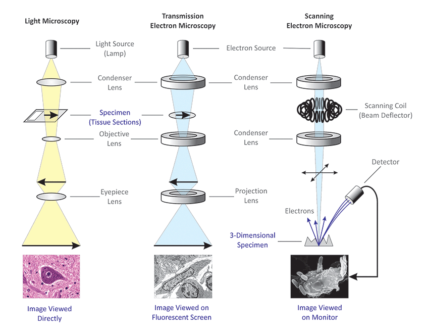
Differences Between Light Microscope And Electron Microscope

Picture Fun Science Scanning Electron Microscope Electron Microscope
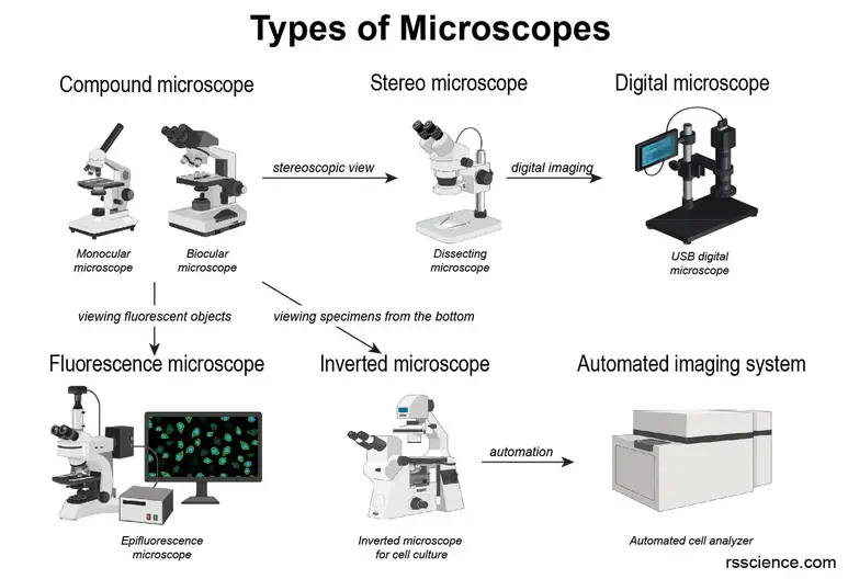
Different Types Of Microscopes Light Microscope Electron Microscope Scanning Probe Microscope Rs Science
Introduction To Electron Microscopy Advanced Microscopy Imaging Facilities The University Of Utah
No comments for "Electron Microscopy Uses Which Type Light for Illumination"
Post a Comment